
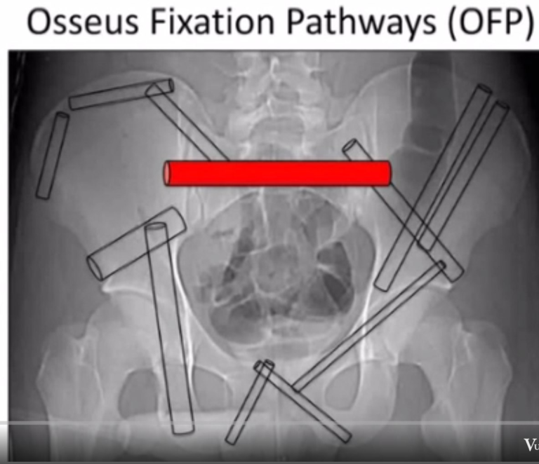
There are seven straight corridors or tunnels in the pelvic bone that allow safe insertion of implants.


Iliosacral Tunnel Through S1
The most commonly used corridor for screw placement. Most affected by sacral dysmorphism. Careful evaluation of X-ray and CT is essential to identify a safe screw path. Intraoperative verification requires interpretation of inlet, outlet, and lateral views and palpation of the drill hole (like probing a pedicle). No X-ray view shows the sacral ala directly — the lateral view shows the iliac condensation that correlates with the alar slope in a normal pelvis.
Iliosacral Tunnel Through S2
Less variable and less risky to neural elements, allowing safe insertion of one transsacral screw from one SI joint to the other based on outlet, inlet, and lateral views.
Supra-acetabular Tunnel
A large column of bone just above the acetabulum and greater sciatic notch, extending from the AIIS to the PSIS. The obturator outlet view (tepee view) shows the axial view of this tunnel; the iliac oblique view shows the length and confirms the screw is above the greater sciatic notch.
This space accepts one or two large screws for pelvic fracture fixation from the front or back, and for lumbopelvic fixation from the back. A screw from the back can be inserted into the S2 ala and advanced across the SI joint into the supra-acetabular tunnel (S2AI screw), making the screw head less prominent posteriorly and aligning it with lumbar pedicle screws to avoid rod bending or connectors.
Anterior Column of the Acetabulum
Extends from the supra-acetabular bone laterally through the superior pubic ramus to the pubic tubercle. The obturator oblique view shows its length and confirms extra-articular implant placement; the inlet view shows anterior or posterior deviation of the drill.
The concavity of the superior pubic ramus medial to the iliopectineal eminence is not easily visualised. Screws can be placed lateral-to-medial or medial-to-lateral.
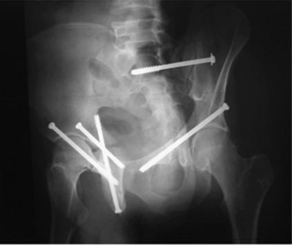
Posterior Column of the Acetabulum
Extends from the pelvic brim to the ischial tuberosity, lying posterior to the acetabulum and anterior to the greater and lesser sciatic notches. Screws can be placed antegrade through the iliac fossa or retrograde from the ischial tuberosity. AP and iliac oblique views are adequate; an oscillating drill-tipped guide wire helps to feel bone better.
Iliac Crest
The bone below the crest is thick and can accept interfragmentary screw fixation of iliac fractures. The crest is thickest over the gluteal pillar, allowing insertion of two to three pins for external fixation.
Inferior Ramus
The corridor from the ischial tuberosity to the pubis along the inferior ramus is rarely used for dedicated implant placement, though screws from anterior pubic plates and posterior ischial plates extend into the inferior ramus to variable lengths.